Computed Tomography, CT or CAT Scans
- Computed tomography creates cross-sectional, multiple-angle images of your body using the lowest possible dose of radiation. The resulting three-dimensional pictures are called tomograms.
- CAT scan testing offers low-dose radiation and high-quality images. Used for diagnostic, biopsy, trauma, vascular and cardiac services, this brief test reveals areas of your body standard X-rays don’t show. With CAT scan, physicians can diagnose certain diseases with speed and precision.
- Our 64-slice CAT scan equipment at both hospitals delivers the most powerful CAT scanning available outside research and teaching facilities. This ultrafast scanning means you only need to hold your breath for short periods of time as the test reconstructs images from any plane or angle.
- CAT scan angiography can scan veins anywhere in the body. This technology is extremely useful for locating blood clots in the lungs and guiding steroid injections into inflamed areas for effective pain management.
- Coronary angiography, a nonsurgical procedure available at Flagstaff Medical Center, can uncover blockages in heart arteries and may be helpful for low-to-medium-risk cardiac patients.
- Low-dose computed Tomography, or LDCT, lung screening is available at our outpatient imaging centers in Cottonwood and Sedona. The U.S. Preventive Services Task Force recommends a lung cancer screening every year for those at high risk, because the sooner lung cancer is caught, the easier it is to treat. An LDCT scan takes only minutes to give you peace of mind.
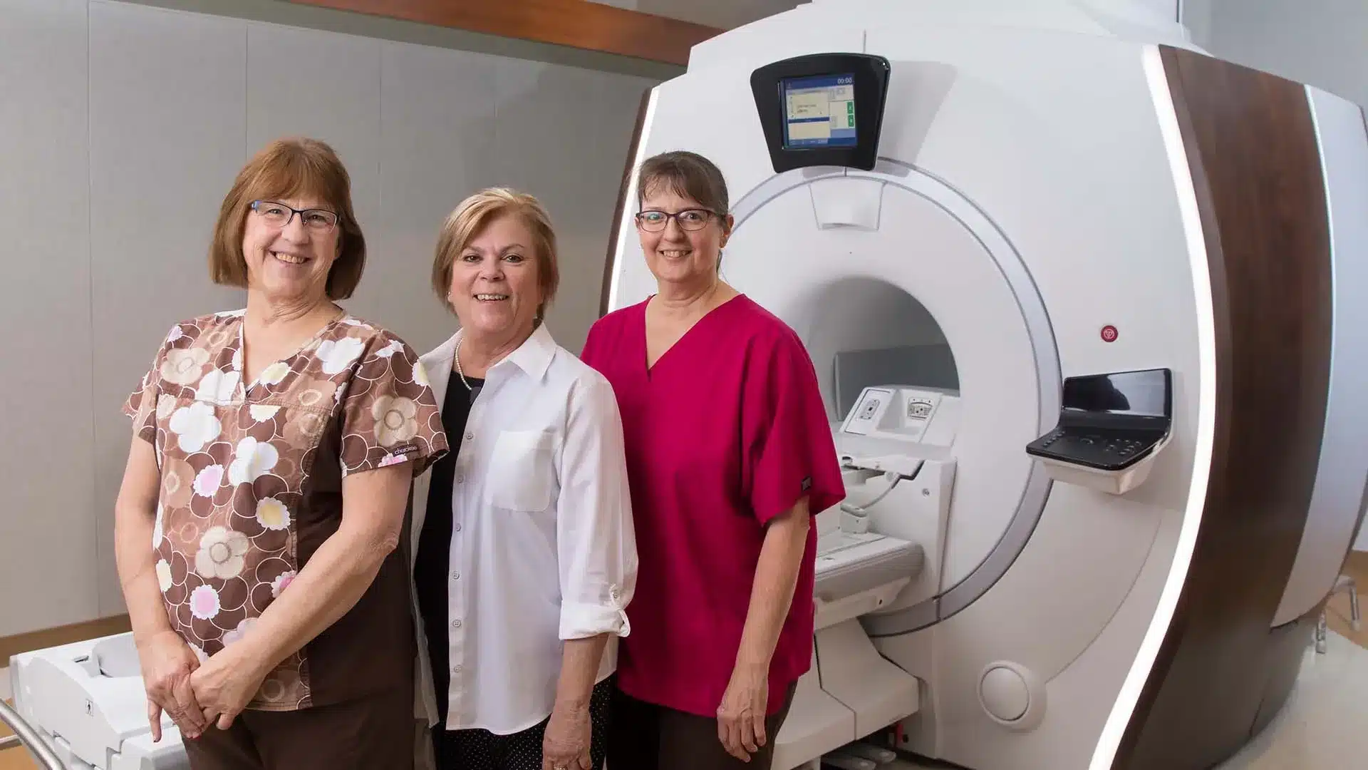
Magnetic Resonance Imaging or MRI
- In magnetic resonance imaging, or MRI, a radio frequency and magnetic field produce high-resolution, two-dimensional, cross-sectional images of bone and soft tissue. This important tool, used in many fields of medicine, can generate a detailed image of any part of the human body. Unlike a CAT scan or X-ray, an MRI doesn’t involve radiation exposure.
- Our imaging professionals use MRI scans to diagnose a variety of conditions, from simple issues like torn ligaments to complex tumors. MRIs are especially useful for examining the brain and spinal cord.
- The Northern Arizona Healthcare Orthopedic Surgery Center uses MRI with 3 Tesla magnetic field strength, while Flagstaff Medical Center and Verde Valley Medical Center use MRI with 1.5 Tesla magnetic field strength. Tesla magnetic field strength refers to the equipment’s ability to capture anatomic detail.
- Northern Arizona Healthcare Medical Imaging Center – Cottonwood offers MRI with 3 Tesla magnetic field strength –the only high-field MRI in the Verde Valley.
Nuclear Medicine
- In nuclear medicine, or “nuc med” for short, small amounts of radioactive material called tracer are directed to a certain organ or part of the body. Test results are accurate, with a minimal amount of radiation exposure. Nuclear medical imaging procedures detect a wide range of conditions, including cancer, heart disease, arthritis and infection.
Positron Emission Tomography or PET
- Positron Emission Tomography, or PET, is the leading diagnostic tool for many types of cancer, coronary heart disease and neurological problems. It combines nuclear medicine technology with X-ray.
- As one of the most advanced, whole-body imaging tools available, PET helps physicians make an earlier diagnosis or determine if a current treatment is working effectively.
- In oncology, PET accurately images multiple organs at the same time to diagnose malignancy and determine if cancer has spread to other parts of the body. PET also looks for evidence of heart attack as well as heart damage that may be reversible if caught in time.
- In neurology, PET is used to investigate brain metabolism in patients with dementia and epilepsy.
- With PET, physicians can look at the smallest chemical and physiological changes to see how a body is functioning. This safe and painless procedure has no known side effects.
Ultrasound
- Ultrasound uses high-frequency sound waves to look organs and structures inside the body. During pregnancy, physicians use ultrasound tests to examine the fetus.
- Ultrasound, unlike X-ray, does not involve radiation exposure. During an ultrasound test, a sonographer or doctor moves a device called a transducer over part of the body. The transducer sends out and receives sound waves that bounce off the tissues inside the body. Images are then created from the sound waves.
- Flagstaff Medical Center offers vascular, complex OB/GYN and abdominal ultrasound exams. Our nationally-registered sonographers perform a variety of procedures, including comprehensive examinations. In April 2017, FMC acquired new, state-of-the-art ultrasound equipment.
- Verde Valley Medical Center uses high-definition ultrasound units with CT sonotechnology for noninvasive vascular exams.
X-Ray/Fluoroscopy
- X-ray technology uses small doses of radiation to create images. Healthcare professionals use these images to look for broken bones and problems in the lungs, abdomen and other areas.
- Different parts of the body will appear light or dark on an X-ray because different body tissues absorb different amounts of radiation. Calcium in bones is the most absorbent, making bones look white on the radiograph. Fat and other soft tissues absorb less and look gray. Air absorbs the least amount, making lungs look black.
- Digital fluoroscopy, or real-time X-ray imaging, offers a variety of imaging equipment with the latest diagnostic capabilities. For most exams, a contrast medium called barium is introduced into the body to highlight specific areas of interest.
- Portable digital imaging enables technologists to conduct X-ray imaging around the clock anywhere in the hospital.
Bone Mineral Densitometry
- Bone mineral densitometry testing measures bone density and is used most often to assess patients for osteoporosis. In a bone density test, X-rays measure how many grams of calcium and other minerals are packed into a segment of bone. The bones most commonly tested are in the spine, hip and forearm.
Interventional Radiology
For interventional radiology procedures, the Northern Arizona Healthcare imaging departments use dedicated special procedures suites. This enables testing for a complete range of interventional procedures, including:
- Vertebroplasty and Kyphoplasty, which treat compressed or fractured vertebrae by placing cement into the vertebra through small, minimally invasive skin incisions under X-ray guidance using fluoroscopy.
- Port-a-cath placements for long-term medication administration, such as chemotherapy.
- Peripherally inserted central catheter, or PICC, placement in the arm. This intravenous line, designed for very ill patients who receive many medications, can be left in place for up to three months. It lessens the number of IV insertions the patient must undergo.
- Drainage placement, which is performed without surgery and involves placing a catheter through the skin and into an organ to drain fluids.
- Arteriogram, to diagnose a blockage or malfunction in the arteries.
- Stent placement, the insertion of a small mesh tube used to treat narrow or weak arteries and improve blood flow.
Digital Mammography
- At Northern Arizona Healthcare Medical Imaging Center – Cottonwood and Northern Arizona Healthcare – Sedona, digital mammography ensures patients have the most advanced technologies and treatments available. A recent medical study proved digital mammograms find cancers traditional mammograms can miss. Digital mammography is especially effective for women who are under 50; pre-menopausal or who have dense breasts.
- All mammography sites have 3-D tomosynthesis.
- According to the American Cancer Society, annual mammography screening is the single most effective method of early detection of breast cancer. Annual mammograms can detect breast cancer when it is most curable – up to two years before a patient or physician can feel any changes in breast tissue.
- These screenings are performed by mammographers – radiologic technologists who are specially trained and registered in the use of this imaging technology.
Stereotactic Breast Biopsy
- In stereotactic breast biopsy, a board-certified radiologist or general surgeon uses a computer to locate and obtain breast tissue samples. Stereotactic technology involves stereo X-rays taken from multiple angles and a special biopsy needle to aspirate, or vacuum, suspicious tissue in a targeted area. Local, instead of general, anesthetic means no hospitalization or prolonged recovery time. Stereotactic breast biopsy is one of the least invasive ways to perform a biopsy and causes minimal disturbance to breast tissue.
- Verde Valley Medical Center uses the very best in breast biopsy equipment to deliver superior image quality, better access and maximum patient comfort.

